First MRI and ultrasound scanning |
|
1. The technology |
2. The human subject |
|
|
|
Magnetic resonance imaging |
|
|
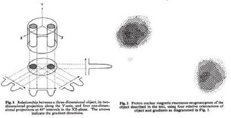
The first MRI image was published in Nature by Paul C. Lauterbur (1929–2007), on 16 March 1973. The image was of his test sample: a 4.2mm diameter test tube containing two water-filled capillary tubes. Lauterbur gave the name 'zeugmatography' to the new imaging technique (although the process was at that time more generally known as Nuclear Magnetic Resonance). [Khened, Kevles, EMRF, Thomas]
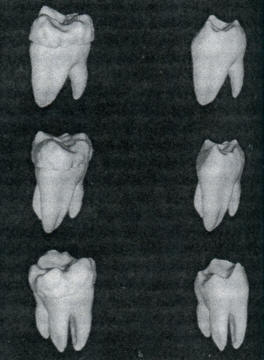
3D volume imaging is achieved by scanning conventional 2D slices of the subject in multiple planes, then assembling these in a 3D spatial presentation. [Khened] In 1995 a team at Caltech led by Pratik Ghosh used a 3D volumetric approach to revealing the structure of a molar removed from the 50-year-old skull of a young woman, in extraordinary detail. This image is reproduced from Kevles. In 2010 scientists Jens Frahm and Axel Haase, at the Max Planck Institute for Biophysical Chemistry in Göttingen, succeeded in significantly reducing the MRI image acquisition time to just one fiftieth of a second. This has enabled the first recordings of MRI motion pictures. Movements of the eye and jaw as well as the bending knee and the beating heart have been filmed. [Max Planck Society] In May 2016 an MRI sound motion picture was uploaded to the internet, showing the baritone Michael Volle singing 'Oh Du, mein holder Abendstern', from Wagner's opera Tannhäuser. The recording was made by the Medical Centre of the University of Freiburg, in Germany.
|
|
|
|
|
Ultrasound scanning |
|
|
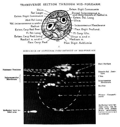
Howry's somagram image of an arm, 1952 |
|
|
Supported by Joseph H. Holmes (1902–1982),
Douglass Howry (1920–1969) produced in 1951, with William Roderic
Bliss and Gerald J. Posakony, both engineers, the 'Immersion tank
ultrasound system', the first 2-dimensional B-mode (or PPI, plan
position indication mode) linear compound scanner. Two-dimensional
cross-sectional images were published in 1952 and 1953, which
convincingly demonstrated that interpretable 2D images of internal
organ structures and pathologies could be obtained with ultrasound.
[History of
Ultrasound in Obstetrics and Gynecology] 3D ultrasound was first developed by Olaf von Ramm and Stephen Smith at Duke University in 1987. [Patent, Duke BME] Often these images are captured rapidly and animated to produce a '4D ultrasound'. |
|
| © 2009–2023 Benjamin S. Beck
|
If you know of any earlier examples, please contact me.
|
|