First invisible radiation photography |
|||
Infrared photography and thermal imaging |
|||
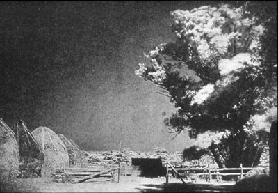
First infrared photograph
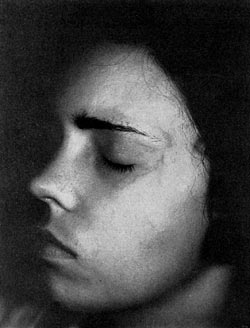
In February 1910 Robert Williams Wood (1868–1955) published the first infrared photographs—landscapes, taken that year on experimental film. One of these landscapes is reproduced above. [Robert W. Wood (February 1910). "A New Departure in Photography". The Century Magazine. The Century Company. 79 (4): 565–572; Pioneers of invisible radiation photography] Colour infrared film was first developed experimentally by two scientists—D.A. Spencer and A. Marriage—working at the Kodak Research Laboratories in England, and further developed at the Kodak lab in Rochester, NY, where a patent was filed on 24 October 1942 and issued on 9 July 1946 in the names of Edwin E. Jelley and Lot S. Wilder (US patent #2,403,722). Military and scientific usage dates from this time. From the early 1960s Eastman Kodak were spooling colour infrared film into 35mm cassettes, from which point it became more accessible to the general public. The process reproduces infrared as red, red as green, and green as blue. [Begleiter, pp12-3; Clark] It seems likely that stereoscopic infrared photographs were made soon after 1910, though I haven't yet found evidence for this. Steven Schwarzman was photographing Stereo Infra-red Landscapes by 1976; his book of that title was published in 1980. A modern example of stereo infrared photography may be found here. Thermal imaging uses long-wavelength infrared, and requires no external light or thermal source. The first thermal imaging camera was developed for the military in Sweden in 1958 by a company called AGA, today known as Flir Systems. [IMVEurope: Seeing without Vision] First infrared photograph of a person
Walter Clark (1899–1991) published an article in 1934 in the Journal of the Biological Photographic Association, on 'Infrared photography'. This included an infrared photograph of a woman's face, which is also reproduced above. [Pioneers of invisible radiation photography]. Steven Schwarzman was photographing infra-red stereo nudes by 1978; his book of them, Bodies of Light, was published in 1981.
|
|||
|
|
|||
Ultra-violet photography |
|||
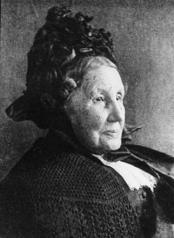
First photograph recording ultra-violet radiation
Hermann Wilhelm Vogel (1834–1898) accidentally discovered the application of short-wavelength photography to medicine in 1864 when he observed black spots on the face of a portrait of a woman—spots that were at the same time hidden to the naked eye. A few days after the photography, however, the woman was found to have smallpox. Films at that time were only sensitive to blue/ultraviolet light, and they would have naturally enhanced the rendition of any skin condition. The significance of Vogel's discovery wasn't recognized at the time. Vogel's 1864 photograph is reproduced above. [Pioneers of invisible radiation photography]. In October 1910 Robert Williams Wood (1868–1955) also published the first reflected ultraviolet photographs—landscapes, and geological and lunar surfaces. First photographs by reflected ultra-violet radiation only
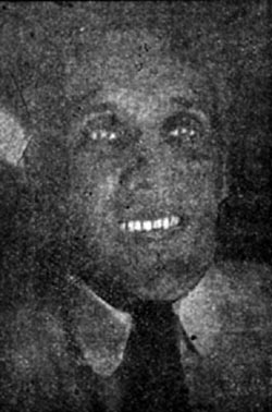
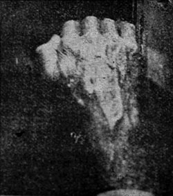
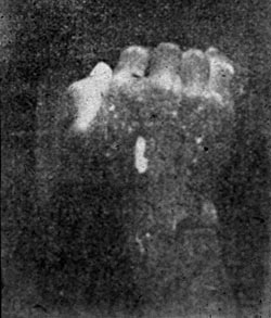
In a paper presented to the French Society of Physics in April 1919, Wood presented three ultraviolet photographs of the human body. The first photograph with the reflected ultraviolet technique showed a face with teeth fluorescing brightly, and he noted how the skin recorded grey. The other two photographs of hands demonstrated the beginnings of a fluorescence technique, which he applied to the back of a hand, recording both the reflected ultraviolet photograph and then his 'phosphorescence' photograph. All three are reproduced above. [Pioneers of invisible radiation photography].
|
|||
|
|
|||
Full-spectrum photography |
|||
| Full-spectrum photography has its roots in spectral imaging, both
multispectral and hyperspectral imaging, which began as early as the
late 1950s / early 1960s as means for geological and military
remote sensing. Full-spectrum photography is a subset of full-spectrum imaging, defined currently among photography enthusiasts as imaging with consumer cameras the full, broad spectrum of a film or camera sensor bandwidth. In practice, specialized broadband / full-spectrum film captures visible and near infrared light, commonly referred to as the 'VNIR'. Modified digital cameras can detect some ultra-violet, all of the visible and much of the near infrared spectrum. Digital cameras normally contain an infrared hot mirror filter that blocks most of the infrared and some of the ultra-violet that would otherwise be detected by the sensor, but replacing the hot mirror or infrared blocking filter with an infrared pass or a wide spectrally transmitting filter allows the camera to detect the wider spectrum light at greater sensitivity. Since 2000 electro-optical engineer David Twede has been developing full-spectrum photography art. Two examples from 2007 may be found on Wikipedia. Examples depicting both men and women can be seen at Surrealmodels. Pioneering work has also been done by Pascal Cotte and his company Lumiere Technology. In October 2004, using a high resolution (240 megapixel) camera, he notably photographed Leonardo's Mona Lisa using a 13-channel acquisition system that employs Melles Griot interference filters in a half-cylinder, covering the visible spectral range, together with one ultra-violet and three infrared filters. [Lumiere Technology, Cotte]
|
|||
|
|
|||
X-ray photography and CT scanning |
|||
|
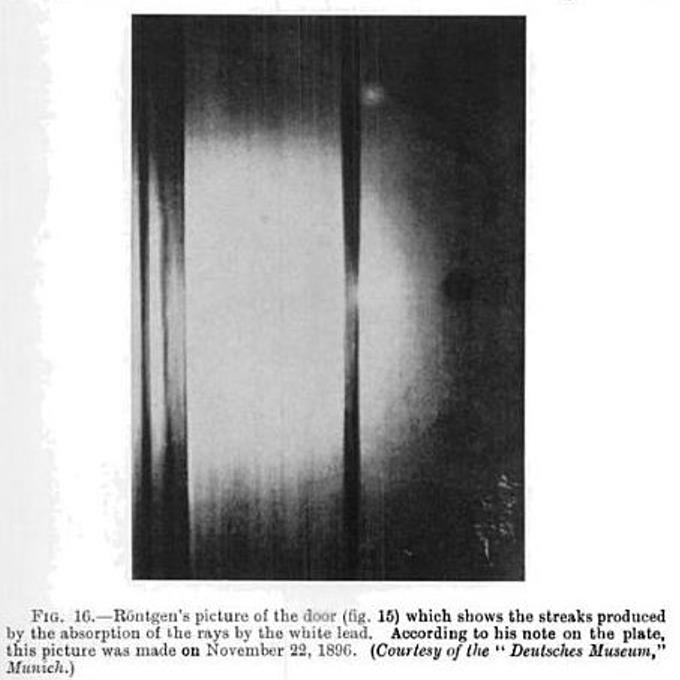
The first X-ray photograph was taken, probably on 22 November 1895, by Wilhelm Conrad Röntgen (1845–1923), of his laboratory door. The image is described as "a photograph made through his laboratory door which registered the varying thickness of stiles and panels, and especially the streaked areas made by brushing on lead-based paint." [Baigrie, Glasser] NB The caption above refers to November 1896; Glasser uses this image, however, to illustrate his account of a demonstration Röntgen gave on 23 January 1896, in which he referred to this door X-ray, so either this is a slip or the image is from a somewhat later repeat of the experiment. [See also Nitske.]
Hand mit Ringen Röntgen took the first X-ray of (part of) a human being on 22 December 1895, photographing the hand of his wife Anna Bertha (Ludwig) Röntgen (1839–1919) (above). (Thomas suggests that he had earlier taken a radiograph of his own hand. He provides no evidence of this, but it's true that Röntgen might have been inclined to experiment on himself before doing so on his wife.
|
|||
|
NB On 22 February 1890 Arthur W. Goodspeed and W.N. Jennings, of the University of Pennsylvania, had accidentally produced an image created by exposure to what were later called X-rays, without (at the time) understanding either the effect or the cause. As may be seen here, it is essentially an X-ray photogram of two coins. See also Penn Medicine. The first full-length, full-size, one-piece X-ray photograph of a complete (living) human being (a woman aged 20) was presented by Kodak in July 1934 at the Century of Progress Exhibition in Chicago. [Kevles] (In 1896 Ludwig Zehnder had exhibited a series of six separate X-ray photographs assembled to depict a whole human skeleton, but in reality they were a composite of parts from three different individuals. See Nitske.) On 1 September 1896 Charles Thurston Holland had taken a whole-body X-ray of a stillborn child; this is reproduced on p69 of Thomas). Stereoscopic X-ray photographs were made as early as the spring of 1896, by Ernst Mach (1838–1916), working with Josef Maria Eder and Eduard Valenta in Vienna: they X-rayed a mouse, viewing the two pictures as diapositives in a mirror stereoscope, which "showed a stereoscopic picture in astonishing relief of the skeleton". Although according to Eder "Roentgen stereoscopy became of unexpected importance in surgery", the more recent view is that it failed to meet medical needs. [Eder, pp384-5; Kevles] A 1920 stereo-pair of the vessels of a foetal torso, by H.C. Orrin, is reproduced on p69 of Thomas). Modern examples are shown at Stereoscopy.com. Autostereoscopic X-ray photographs were produced experimentally by Théodore Guilloz (1868–1916) in 1904, using the line-screen process. He exhibited stereoscopic line-screen X-rays of "nails, screws, metal objects in complicated arrangements," and "the hand and the wrist of a live adult" at the Société de Biologie that year. [Timby, p27] In 1897 John Macintyre (1859–1928), who set up the world's first radiology department at the Glasgow Royal Infirmary, produced the first experimental roentgencinematographs (i.e. X-ray motion pictures), filming small subjects, such as a frog's leg, directly (1:1). [Tosi, Kevles] The frog's leg, and other X-ray motion film by Macintyre, may be seen on the Origins of Scientific Cinematography DVD, as well as at the Scottish Screen Archive, where it is dated to 1896. Computed tomography (CT) is a medical imaging method employing tomography. Digital geometry processing is used to generate a 3D image of the inside of an object from a large series of two-dimensional X-ray images taken around a single axis of rotation. As early as 1965 David Kuhl had achieved the first transmission section scan of a living human thorax, by sending radioactive beams through the subject. [Kevles, which reproduces the image] The first CT images, from 1969 or 1970, were of inanimate subjects, specifically Perspex 'phantoms' of varying complexity, and later of a human brain section in formalin, in a Perspex box. Readings employed a scintillation scanner to count gamma ray photons; it took nine days to take a picture and 15 minutes of computer time to reconstruct it. A photograph of an example of a CT image of a group of Perspex phantoms is reproduced in Thomas.
[Maier A, Steidl S, Christlein V, et al., es. (2018) Medical Imaging Systems: An Introductory Guide] On 1 October 1971, CT scanning was introduced into medical practice with a successful scan on a patient at Atkinson Morley Hospital in Wimbledon, London—a woman of 41, with a suspected cystic tumour in the left frontal lobe (confirmed by the scan). The initial CT scanner used an 80 x 80 matrix for image display, and the first images (of two contiguous slices) were obtained as Polaroid photographs of the monitor screen. [BJR, Kevles] This image was 2-dimensional, of course. According to Thomas, the scanning time was four minutes per slice, with a slice thickness of just over 1 cm. As no computer was attached to the scanner the data was saved onto magnetic tape and driven to Hayes to be analysed by EMI on an ICL 1905 mainframe computer; the images took 20 minutes to construct. The original Polaroid is inscribed on the back by Godfrey Hounsfield, "original 1st PATIENT SCANNED". [Thomas]. However, Godfrey Hounsfield (1919–2004), the inventor, had previously built a prototype head scanner and tested it first on a preserved human brain, then on a fresh cow brain from a butcher's shop, and later on himself. Whole body CT scanning was first achieved by Louis Kreel (1925–2019) in 1975, at Northwick Park Hospital in Harrow, at which time carcinoma of the pancreas was diagnosed. In 2001 the Massachusetts-based Actuality Systems introduced the prototype of its Perspecta Spatial 3-D system, which is a volumetric, autostereoscopic display system, chiefly used in the medical market. Perspecta allows users to view moving objects from any angle with the unaided eye, simply by walking around them as you would if you were looking at real 3D objects. Perspecta consists of a rotating round white polymer screen resting on a box containing software, hardware, and an optical system. 25cm-diameter images appearing to float inside a clear sphere (field of view 360º horizontal, 270º vertical) are generated by slices of successive 2D images, rapidly projected one after another onto the screen, creating an illusion of a real 3D image. Perspecta Display 1.9 allows for full-colour imagery, and objects in motion. This is particularly effective with CT imagery, for example of breathing lungs. The Perspecta system was featured as a medical investigation tool in the CSI: New York autopsy lab in Season 4, Episode 1, broadcast on 26 September 2007. Actuality Systems was dissolved at the end of 2009, and intellectual property rights are now held by Optics for Hire. [Actuality Systems, Genuth]
|
|||
|
|
|||
Neutron tomography |
|||
|
Neutron tomography has been available since 1997 at the Neutron Transmission Radiography Station (NEUTRA) of the Paul-Scherrer-Institute (PSI) in Villigen, Switzerland. The technique is similar to X-ray CT but, unlike X-rays, neutrons interact significantly with some light materials (e.g. hydrogenous substances) and penetrate heavy materials with ease. It is therefore complementary to X-ray tomography. In the PSI a particle accelerator directs a proton beam at a lead target, generating neutrons. The neutrons are then slowed down to thermal energy and the rays concentrated to an almost parallel 35 cm beam. This beam is transmitted through the object and recorded by a plane position sensitive detector. For tomographic images, the object is placed on a rotating table and turned in small angular steps through 180° while a camera system takes several projections. From the single projections, slices perpendicular to the rotation axis are reconstructed by a tomographic reconstruction algorithm. The slices are then collected in an image stack that can be visualized using 3D rendering software. [Schwarz et al., NEUTRA] The technique is suitable for the visualization of the inner structure of industrial, biological, and geological structures; in particular it has been found useful in exposing the internal structures of fossils. [MNRC, Schwarz et al.]
|
|||
|
|
|||
Gamma ray tomography |
|||
| PET (Positive Emission Tomography) and SPECT (Single Photon Emission
Computed Tomography) both employ gamma radiation to create images.
However both require the introduction of radioisotopes to the
subject's body, which for present purposes really puts them beyond
the intended scope of these pages. The first gamma camera (also called the scintillation or Anger camera) was developed in 1957 by Hal Anger (1920–2005). [Journal of Nuclear Medicine]
|
|||
|
|
|||
Terahertz imaging |
|||
| Terahertz radiation falls between microwaves and infrared in the
electromagnetic spectrum. Imaging applications identified so
far range from quality control to security screening, but there is
potential for much wider use. The first work in terahertz imaging was published in the mid-1970s by T.S. Hartwick et al., but none of the published images seem to be available online without a subscription. [T.S. Hartwick, D.T. Hodges, D.H. Baker, and F.B. Foote (1976) 'Far infrared imagery', Applied Optics 15:1919–22] This technology was workable in the lab, but proved too complicated to inspire much further effort on the same lines. It wasn't until 1995 that a new technique, terahertz time-domain spectroscopy (THz-TDS), developed by B.B. Hu and Martin C. Nuss, awakened real interest and significant subsequent development. [B.B. Hu and M.C. Nuss (1995) 'Imaging with terahertz waves', Optics Letters 20:1716–19]
[Imaging with terahertz radiation, Teraherz imaging comes into view]
|
|||
|
© 2010–2024 Benjamin S. Beck
|
If you know of any suitable examples, please contact me.
|
||

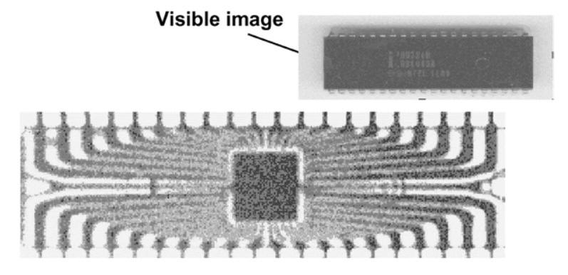 One of the first images acquired
using THz-TDS, from Hu and Nuss (1995). It shows a
transmission image of a semiconductor integrated
circuit, through the black epoxy package, which is
transparent to terahertz radiation. The circuitry and
the central semiconductor wafer are clearly seen. The
inset shows an optical view of the same object.
One of the first images acquired
using THz-TDS, from Hu and Nuss (1995). It shows a
transmission image of a semiconductor integrated
circuit, through the black epoxy package, which is
transparent to terahertz radiation. The circuitry and
the central semiconductor wafer are clearly seen. The
inset shows an optical view of the same object.